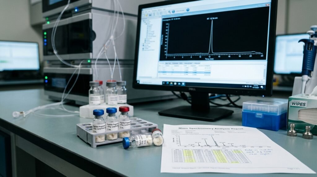

The melanocortin system has long been investigated for its broad control over pigmentation, energy metabolism, and immunomodulatory cascades. However, full-length alpha-melanocyte-stimulating hormone ($\alpha$-MSH), a 13-amino-acid neuroimmunomodulatory peptide, presents experimental challenges in non-pigmentary studies due to its potent stimulation of melanogenesis via melanocortin 1 receptors (MC1R).
To isolate the peptide’s anti-inflammatory characteristics from pigmentary effects, researchers turned to its C-terminal sequence: the tripeptide KPV (Lysine-Proline-Valine).
KPV is a naturally derived C-terminal tripeptide fragment ($\alpha\text{-MSH}_{11\text{–}13}$):
$$\text{H-Lys-Pro-Val-OH}$$
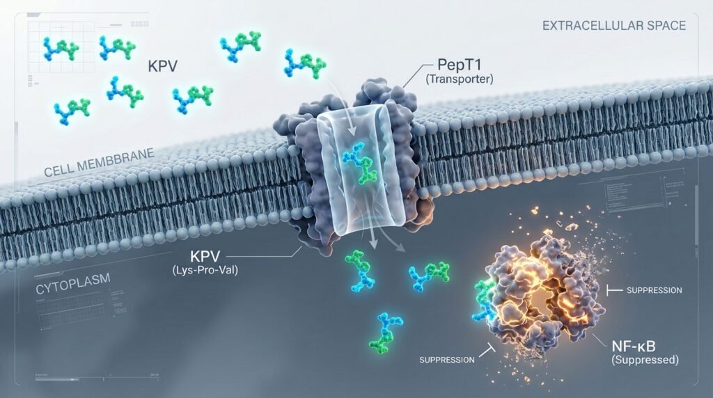
With a low molecular weight ($M_w \approx 384.5\text{ g/mol}$), KPV does not rely exclusively on traditional receptor-mediated endocytosis. Instead, cellular studies demonstrate that KPV utilizes the peptide transporter 1 (PepT1) system:
The central scientific interest in KPV centers around its targeted suppression of the nuclear factor kappa B (NF-$\kappa$B) transcription factor cascade.
Pro-Inflammatory Stimulus (LPS / TNF-α)
                     │
                     ▼
             IκB Phosphorylation
                     │
         [ KPV Inhibitory Blockade ]
                     │
                     ▼
          NF-κB Nuclear Translocation Blocked
                     │
         ┌───────────┴───────────┐
         ▼                       ▼
Downregulated IL-1β, IL-6    Suppressed iNOS & COX-2
Under baseline inflammatory stimulation (e.g., exposure to lipopolysaccharide or TNF-$\alpha$), the inhibitor protein I$\kappa$B$\alpha$ is phosphorylated by IKK and targeted for proteasomal degradation. This event liberates the NF-$\kappa$B p50/p65 heterodimer to enter the nucleus. In epithelial and macrophage cell lines, intracellular KPV prevents I$\kappa$B$\alpha$ degradation, effectively trapping NF-$\kappa$B in an inactive state within the cytosol.
By suppressing NF-$\kappa$B nuclear translocation, KPV-treated cultures show significant decreases in downstream effector transcripts:
Preclinical microbiological investigations have documented direct, sequence-specific antimicrobial actions against Staphylococcus aureus and Candida albicans, suggesting that KPV disrupts microbial membrane potential while dampening host inflammatory tissue damage.
Due to its tripeptide size and proline residue, KPV exhibits high aqueous stability:
Disclaimer: Strictly for scientific laboratory experimentation and in-vitro research. Not intended for therapeutic use.

Augue Sed viverra nulla Interdum mia bibendum velit sapien usop scelerisqu ictum quam tincidunt nec feugiat augue tincidunt Etiam pretium diam rhoncus. gida turpis cursus. Nuncsed fringilla tortor iaculis eget tincidunt accumsan ullamcorper.